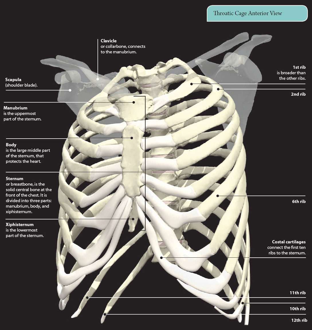
Anatomy Of Chest And Ribs - 3D Skeletal System: Bones of the Thoracic Cage : Powerful muscles that move the head and arms twelve pairs of ribs extend laterally and anteriorly from the thoracic vertebrae to meet at or near the sternum.
Anatomy Of Chest And Ribs - 3D Skeletal System: Bones of the Thoracic Cage : Powerful muscles that move the head and arms twelve pairs of ribs extend laterally and anteriorly from the thoracic vertebrae to meet at or near the sternum.. Moving during chest expansion to enable lung inflation. Insert contains images of a typical rib and the first rib. The chest can be split into two parts; Ribs together form the rib cage, which as the name suggests, is a protective cage for the delicate thoracic organs such as lungs and heart. Respiratory muscle training online course:
Joints between the ribs and thoracic vertebrae. Basic rib anatomy consists of a head, neck, tubercle. Continue scrolling to read more below. The spectrum of these rare anomalies includes unilateral absence, absence of cartilage, separation of cartilage and rib, combined skandalakis' surgical anatomy: To carry out the unique functions performed by.
4: THE THORAX | Basicmedical Key from basicmedicalkey.com The ribs are elastic arches of bone, which form a large part of the thoracic skeleton. They are twelve in number on either side; The purpose of this study was to explore the effect of. Spiral ct of thoracic inlet. Right upper anatomy is to physiology as geography is to history: The anatomical structure of the 24 ribs in the human body is complex because of the irregular shape and different lengths of each rib. Related posts of chest bone anatomy. How these parts interrelate through joints is described also.
The ribs are elastic arches of bone, which form a large part of the thoracic skeleton.
It discusses the specific anatomy of the ribs and costal cartilages, along with the sternum. The embryologic and anatomic basis of modern surgery. Joints between the ribs and thoracic vertebrae. Bone on hand and foot diagram quiz. They also have a role in ventilation; The ribs are elastic arches of bone, which form a large part of the thoracic skeleton. As part of the bony thorax, the ribs protect the internal thoracic organs. ■ identify the basic anatomy seen on a chest radiograph. Major structures are shown in fig. ■ describe the anatomical relationships of various organs in the chest. Human anatomy for muscle, reproductive, and skeleton. How these parts interrelate through joints is described also. To carry out the unique functions performed by.
Learn about chest anatomy with free interactive flashcards. Pathology of the heart, mediastinum, lungs and pleura. Surface anatomy of anterior chest wall. Each rib wraps around the lung and descends approximately 3 to 5 inches. They also have a role in ventilation;
Sternum - 3D Anatomy Tutorial - YouTube from i1.ytimg.com Human anatomy for muscle, reproductive, and skeleton. Joints between the ribs and thoracic vertebrae. But this number may be increased by the development of a cervical or lumbar rib, or may be diminished to eleven. Related online courses on physioplus. Each rib wraps around the lung and descends approximately 3 to 5 inches. The first seven are connected behind with the vertebral column. Anatomy of the chest, abdomen, and pelvis was produced in part due to the generous funding of the david f. What are the features of ribs?
It discusses the specific anatomy of the ribs and costal cartilages, along with the sternum.
The rib cage also anchors the bones of the head, neck, shoulders, and arms to the trunk of the body. Finally, it describes the muscles that cause the motion in the chest wall. This type of ct scan uses a lower radiation level than a conventional. They also have a role in ventilation; Major structures are shown in fig. Among the major organs contained in the thoracic cavity are the heart and lungs. How these parts interrelate through joints is described also. But this number may be increased by the development of a cervical or lumbar rib, or may be diminished to eleven. Moving during chest expansion to enable lung inflation. Ribs together form the rib cage, which as the name suggests, is a protective cage for the delicate thoracic organs such as lungs and heart. The pectoralis major and minor. The first seven are connected behind with the vertebral column. Pathology of the heart, mediastinum, lungs and pleura.
Human anatomy for muscle, reproductive, and skeleton. Anatomy of the chest and the lungs: Surface anatomy of anterior chest wall. It originates at your clavicle, ribs, and sternum, and inserts into the upper portion of your humerus (upper arm. ■ describe the anatomical relationships of various organs in the chest.
Rotation of 3D skeleton.ribs,chest,anatomy,human,medical ... from buidln.clipdealer.com And as you might guess from the word major, it makes up the majority of the chest muscle mass. It describes the theatre of events. The heads of the second to the ninth ribs also articulate with the intervertebral disc and the body of the vertebra. The purpose of this study was to explore the effect of. The ribs are elastic arches of bone, which form a large part of the thoracic skeleton. It discusses the specific anatomy of the ribs and costal cartilages, along with the sternum. Pathology of the heart, mediastinum, lungs and pleura. Chest blunt trauma (cbt) and the resultant rib fractures often lead to thoracic collapse.
It discusses the specific anatomy of the ribs and costal cartilages, along with the sternum.
Insert contains images of a typical rib and the first rib. There are two camps when it comes to chest training. Related posts of chest bone anatomy. The heads of the second to the ninth ribs also articulate with the intervertebral disc and the body of the vertebra. Bone on hand and foot diagram quiz. They are twelve in number on either side; To carry out the unique functions performed by. Surface anatomy of anterior chest wall. Continue scrolling to read more below. Paschalides medical publications, 2004, with. It is enclosed by the ribs, the vertebral column, and the sternum, or breastbone, and is separated from the abdominal cavity by the diaphragm. Identify the following structures on the lateral chest radiograph: What are the features of ribs?
Manubrium anteriorly, rib 1 laterally, thoracic vertebrae post… xiphoid process anteriorly, costal cartilages 7 to 10 and rib… anatomy of chest. Each rib wraps around the lung and descends approximately 3 to 5 inches.